Latest News & Events
Glenbrook LabScope™ Reveals Highly Detailed In-Vivo Images for Orthopaedic Researchers
 April 2010
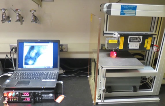
(RANDOLPH, NJ) – When researchers at the Hospital for Special Surgery in Manhattan needed to look inside a rat’s knee to observe the reconstruction and healing process of a new surgical procedure, they came to Glenbrook Technologies in Randolph, NJ to use the LabScope, Glenbrook’s patented magnification fluoroscopy technology.
LabScope technology displays anatomical details in high resolution – up to 15 line pairs per millimeter – and up to 25X magnification, using low-dose radiation that requires minimal operator protection. The compact, low-cost system displayed the rat’s patella in highly detailed still images and motion studies that were saved to memory for future reference. Subsequently, Glenbrook lent a LabScope to HSS for the staff to use to complete its research studies. Since then, the Center for Disease Control in Atlanta has purchased two LabScopes. The potential orthopaedic research applications of the LabScope cover a wide variety of small animal imaging studies.
Glenbrook Technologies, founded in 1983, has developed numerous x-ray technologies for the animal research industry as well as the electronic, medical device and security industries. In 2009 the company received both a Thomas Alva Edison Patent Award from the Research and Development Council of New Jersey and an Invention Advancement Award from the New Jersey Inventors Hall of Fame.
For more information about Glenbrook Technologies’ innovations in x-ray imaging technology, call
973-361-8866 or visit www.glenbrooktech.com.
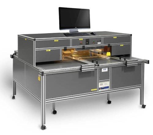
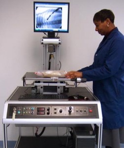
Extraordinarily magnified fluoroscopic detail and small system footprint achieved with patented technology
Minimal protective garments required
Images and motion videos saved to memory
For information about Glenbrook Technologies’ innovations in x-ray imaging technology,
call 973-361-8866